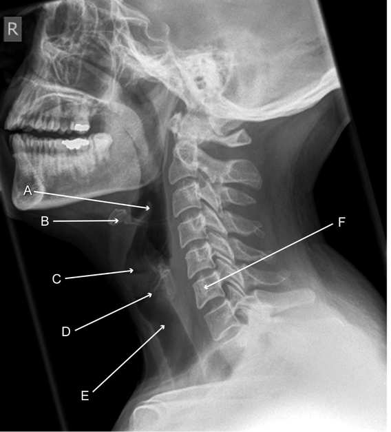
- Epiglottis. Composed of fibrocartilage, it projects obliquely behind the base of the tongue, in front of the entrance to the larynx. The space between the base of tongue and epiglottis is termed the vallecula and is a potential site for foreign bodies
- Hyoid bone. This horseshoe shaped bone in the anterior midline of the neck lies at the level of the third cervical vertebral body
- Laryngeal ventricle. This is the space between the true and false vocal cords. It is seen as a dark line on plain film, the lower border of which indicates the level of the true cords
- Calcified cricoid cartilage. The cricoid is the only complete cartilaginous ring of the trachea. It sits inferior to the thyroid cartilage, and often becomes calcified. The degree of calcification is proportional to age and calcification is more common in men than in women.1 Calcified cricoid cartilage is often mistaken for an ingested foreign body
- Lumen of the trachea. Lying anterior to the oesophagus, the trachea acts as a useful anatomical marker to assess whether a structure lies within the oesophagus
- Sixth cervical vertebra. This is an important landmark when interpreting a lateral soft tissue neck radiograph because the cricoid cartilage lies between the level of the fifth and sixth cervical vertebrae. This indicates the level of cricoid cartilage and cricopharyngeus muscle, the most common site of foreign body entrapment
Source: British Medical Journal
BMJ 2013;347:f6186