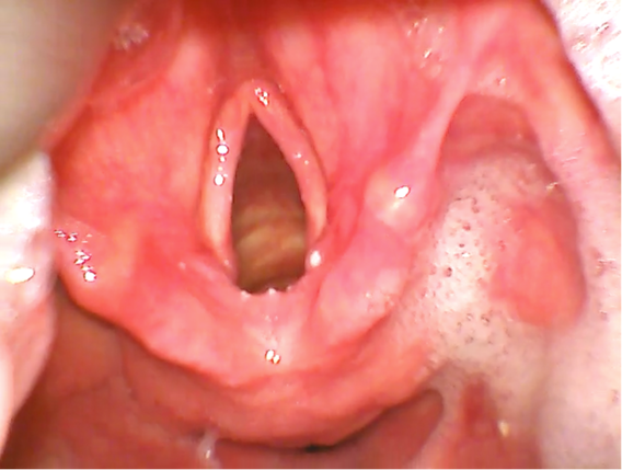
The Cormack–Lehane (CL) classification is a grading system commonly used to describe laryngeal view during direct laryngoscopy. It referred to the best view obtained, i.e. with or without manipulation of the larynx. First published in 1984, this classification was described in order to teach trainees in obstetric general anaesthesia as a means of simulating difficult tracheal intubation and not as a grading system for everyday recording of the view at laryngoscopy.
The incidence of difficult intubation in surgical patients undergoing general anesthesia is estimated to be approximately 1–18%, whereas that of failure to intubate is 0.05–0.35%
Although there are many airway assessments available for laryngoscopy such as Mallampati classification, mouth opening, upper lip bite test, there is still no single test with 100% sensitivity and specificity to predict difficult laryngoscopy and intubation.
Did you know?
In 2008, an interview was conducted to assess the interpretation of the Cormack and Lehane view at the European Society of Anaesthesiologists congress. Although 89% claimed to know a classification of laryngeal view during laryngoscopy, only 53% were able to name the classification and only 25% could describe all four grades correctly.
The ‘Fremantle Score’ is a new scoring system introduced to describe the view, ease and device used for videoscopic intubation.
Whenever we refer to the laryngoscopy view, we will refer to the Cormack and Lehane grading classification in the intubation videos.
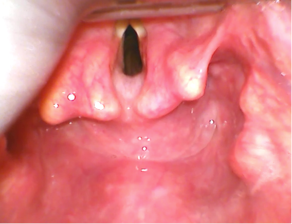
Grade 1 view
Most of the glottic opening can be seen.
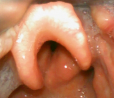
Grade 2 view
Only the posterior portion of the glottis or only arytenoid cartilages are visible.
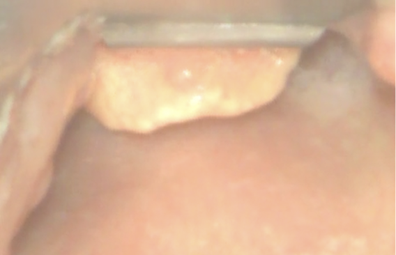
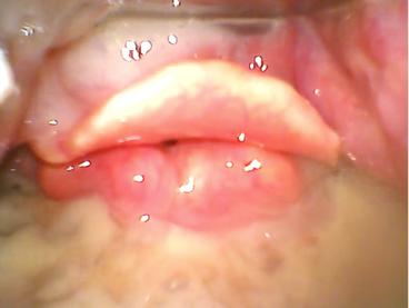
Grade 3 view
Only the epiglottis but no portion of the glottis is visible.
In grade 4 view , neither the glottis nor the epiglottis can be seen.
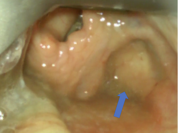
Image captured during laryngoscopy demonstrates pyriform sinus on the right (arrow). It is a common location for impacted food.
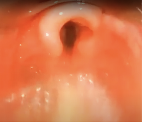
Image captured during laryngoscopy showing normal larynx anatomy in an infant. Note that the epiglottis is narrow, long, floppy and omega shaped. The vocal cords are shorter.
Other characteristics of pediatric larynx:
- Shape is conical compared to the cylindrical shape in adults.
- The larynx in infants and young children is located more anterior and superior compared with the adult’s.
- The narrowest portion of the pediatric airway was previously believed to be below the glottis at the level of the cricoid cartilage. However recent MRI studies report the narrowest portion of the pediatric airway to be at the level of vocal cords similar to adults.
The angle of tracheal bifurcation in infants and children is found to be approximately 80-110°. Hence the chances for mainstem intubation is equal on right and left. However, in adults the right bronchus is straighter (20°to right of midline) compared to the left bronchus (>35°to left of midline) increasing the chances for right mainstem intubation if the endotracheal tube is advanced beyond the carina.
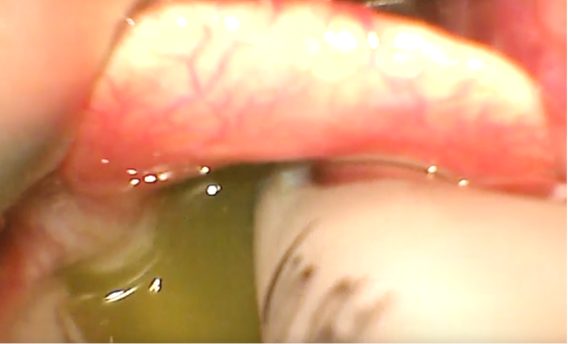
Bile seen in larynx during an intubation.
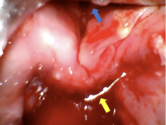
A patient with active gastrointestinal bleeding (shown by yellow arrow). Blue arrow points towards the larynx. Blood in airway can obscure vision especially when a videolaryngoscope or fiberoptic bronchoscope is used.
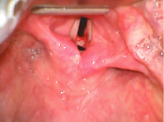
Pathological Conditions:
Vocal cord web